Masson Trichrome Kit
Connective Tissue Stain — Collagen Differentiation
Masson Trichrome Kit
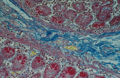
Masson's Trichrome is the most widely used connective tissue stain in histopathology. This three-dye technique differentially stains collagen fibers, muscle, cytoplasm, and nuclei in distinct colors. Collagen stains blue or green (depending on formulation), cytoplasm and muscle stain red or pink, and nuclei stain dark brown or black. Essential for evaluating fibrosis in liver biopsies, studying renal pathology, diagnosing muscular dystrophy, and assessing cardiac tissue changes. The clear differentiation makes it invaluable for quantifying collagen deposition in chronic diseases.
Masson Trichrome Results
Collagen Fibers
Stain blue (aniline blue formulation) or green (light green formulation). Type I and III collagen clearly visible.
Cytoplasm & Muscle
Stain red with Biebrich scarlet or acid fuchsin. Distinguishes muscle from connective tissue.
Nuclei
Stain dark brown to black with Weigert's iron hematoxylin. Chromatin detail preserved.
Fibrin
Stains bright red, useful for identifying thrombi and fibrin deposits in tissue sections.
Complementary Stains
Need Special Stain Kits?
Contact our team for specifications, protocols, and availability.