Gordon & Sweets Kit
Reticulum Fiber Stain — Silver Impregnation
Gordon & Sweets Reticulum Kit
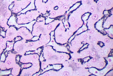
The Gordon & Sweets staining method is the classical silver impregnation technique for demonstrating reticulum fibers in tissue sections. This kit impregnates reticular fibers with silver salts, making them appear black against a pale background. Essential for evaluating liver architecture, identifying cirrhosis, detecting early fibrosis, and studying lymphoid tissue framework. The reticulum stain is critical in hepatopathology, renal pathology, and hematopathology for assessing the integrity of connective tissue support structures.
Gordon & Sweets Results
Reticulum Fibers
Stain black due to silver impregnation. Fine branching fibers form supportive network around cells.
Nuclei
Stain gray to black depending on hematoxylin counterstain used. Provides cellular detail.
Background
Cytoplasm and collagen appear pale pink or gray, providing contrast to black reticulum fibers.
Clinical Applications
Essential for liver biopsy evaluation, lymphoma diagnosis, and assessing fibrosis in chronic diseases.
Connective Tissue Stains
Need Special Stain Kits?
Contact our team for specifications, protocols, and availability.