Vangieson Kit
Elastic Fiber Stain — Connective Tissue
Vangieson Elastic Fiber Kit
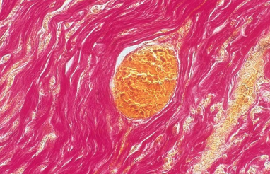
The Vangieson staining method is a specialized technique for demonstrating elastic fibers in tissue sections. This stain is particularly valuable for evaluating blood vessel walls, studying skin biopsies, and examining connective tissue disorders. Elastic fibers stain dark purple or black against a contrasting background, making them easily distinguishable from collagen and other connective tissue components. The Vangieson method is essential in cardiovascular pathology for assessing arterial wall integrity, in dermatopathology for evaluating solar elastosis and pseudoxanthoma elasticum, and in pulmonary pathology for studying emphysema and elastic tissue changes.
Vangieson Staining Results
Elastic Fibers
Stain dark purple to black. Resorcin-fuchsin has high affinity for elastin, revealing delicate fiber networks in tissues.
Collagen
Stains pink or red with van Gieson counterstain. Clear differentiation from elastic fibers aids tissue evaluation.
Nuclei
Stain blue with optional hematoxylin counterstain. Provides cellular detail within connective tissue framework.
Clinical Applications
Essential for arterial disease evaluation, skin pathology (elastosis), and connective tissue genetic disorders.
Connective Tissue Stains
Need Special Stain Kits?
Contact our team for specifications, protocols, and availability.